
Diabetic Eye Care
The primary cause of diabetic retinopathy is diabetes—a condition in which the levels of glucose (sugar) in the blood are too high. Elevated sugar levels from diabetes can damage the small blood vessels that nourish the retina and may, in some cases, block them completely.
When damaged blood vessels leak fluid into the retina it results in a condition known as diabetic macular edema which causes swelling in the center part of the eye (macula) that provides the sharp vision needed for reading and recognizing faces.
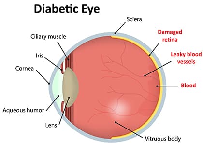
Prolonged damage to the small blood vessels in the retina results in poor circulation to the retina and macula prompting the development of growth factors that cause new abnormal blood vessels (neovascularization) and scar tissue to grow on the surface of the retina. This stage of the disease is known as proliferative diabetic retinopathy (PDR).
New vessels may bleed into the middle of the eye, cause scar tissue formation, pull on the retina, cause retinal detachment, or may cause high pressure and pain if the blood vessels grow on the iris, clogging the drainage system of the eye—all of this can cause vision loss.
The best way to diagnose diabetic retinopathy is a dilated eye exam. During this exam, the physician places drops in the eyes to make the pupils dilate (open widely) to allow a better view of the inside of the eye, especially the retinal tissue.
- Swelling in the retina that threatens vision (diabetic macular edema)
- Evidence of poor retina blood vessel circulation (retinal ischemia)
- Abnormal blood vessels that may predict an increased risk of developing new blood vessels
- New blood vessels or scar tissue on the surface of the retina (proliferative diabetic retinopathy)
Regular dilated eye exams by a Retina specialist are important, especially for those who are at a higher risk for diabetic retinopathy or diabetes. If you are over age 50, an exam every 1 to 2 years is a good idea so the physician can look for signs of diabetes or diabetic retinopathy before any vision loss has occurred.
In addition to this exam, physicians use other tests to detect and manage diabetic retinopathy:
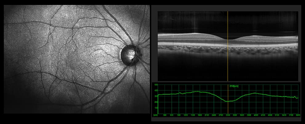
An optical coherence tomography (OCT) test provides highly detailed cross-sectional images of the retina that show its thickness, helping determine whether fluid has leaked into retinal tissue.
The physician may take fundus photographs of the back of the eye to help detect and document diabetic retinopathy. These photos make it easier for the physician to monitor the disease on follow-up visits to determine if it is worsening.
To evaluate retina blood vessel circulation, the physician may conduct a retinal photography test called fluorescein angiography (FA). After dilating the pupils, the physician will inject a dye into the patient’s arm. The dye then circulates through the eyes and works like a food coloring; however, it does not affect the kidneys and is unlike the dye that is used with MRIs and CAT scans.
As the dye circulates, the physician takes pictures of the retina to accurately detect blood vessels that are closed, damaged, or leaking fluid. The pictures are black and white to help the doctor detect these changes more easily, but the process is not the same as having an x-ray. Prior to examination, ask your physician to discuss the risks and benefits of obtaining these images.
With proper examinations, diabetic retinopathy can be detected before vision loss begins. If the physician detects signs of diabetic retinopathy, she/ he will determine how frequently follow-up examinations will be required to detect changes that would require treatment.
Treatment and prognosis
There are many approved treatments for diabetic retinopathy, including intravitreal injections (small injections of medications into the middle cavity of the eye), laser treatments, and vitreous and retina surgery. These procedures can be done in an office or hospital setting to prevent, treat, or reverse damage from diabetes in the retina.
Research has shown that eye injections often result in better vision than laser treatment alone for patients with diabetic macular edema. The key to these treatments is their ability to block vascular endothelial growth factor (VEGF), a chemical signal that stimulates leakage and abnormal blood vessel growth. Repeated doses of anti-VEGF medications may be needed to prevent blood vessels from leaking fluid and causing vision loss.
Even if not all vision loss from diabetic retinopathy can be prevented or treated, patients usually are able to find resources to help them live with diminished vision. If you have been diagnosed with diabetic retinopathy or diabetes and have vision loss that cannot be reversed, a retina specialist can help you find access to rehabilitation with a variety of tools to make everyday living with this disease a little bit easier. A retina specialist can also help connect you with others who have similar limitations.